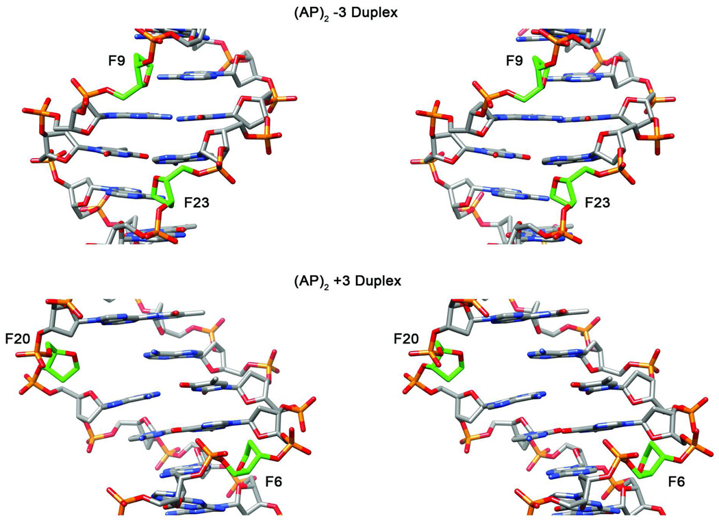
Figure 8.
Expanded cross eye stereo view (63, 64) of the (AP)2−3 cluster seen with the minor groove prominent and the (AP)2+3 cluster seen with the major groove prominent, when the Watson-Crick alignments at the lesion site restraints are enforced during MD. AP residues are colored green. In the case of the (AP)2−3 duplex, the AP residues are closer across the minor groove while they are farther apart across the major groove in the (AP)2+3 duplex.

