Fig. 3.
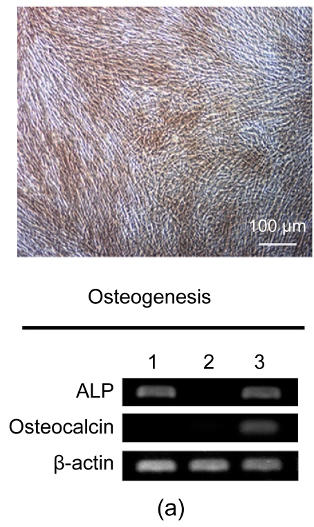
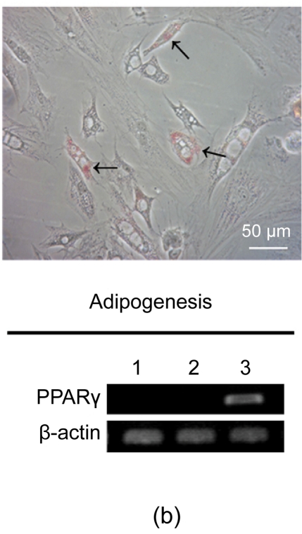
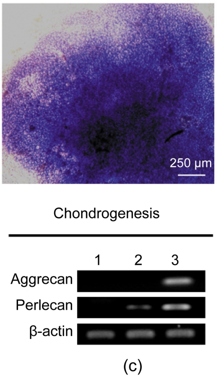
Osteogenic, adipogenic, and chondrogenic differentiations of hESC-MSCs
(a) After three weeks, osteogenesis was demonstrated by calcium deposition in the matrix visualized with von Kossa staining and increased expression of ALP and Osteocalcin; (b) Lipid droplets (arrows) were detectable by oil-red O staining after two weeks of adipocytic induction and PPARγ, a marker of adipocytic differentiation, was detected by RT-PCR; (c) While after three weeks of induction, chondrogenic differentiation of hESC-MSCs was achieved. More than 80% of all cells stained positively with toluidine blue. The expressions of chondrogenic genes, aggrecan and perlecan, were confirmed by RT-PCR. Lane 1: hESCs; Lane 2: hESC-MSCs; Lane 3: Induced cells


