Figure 2.
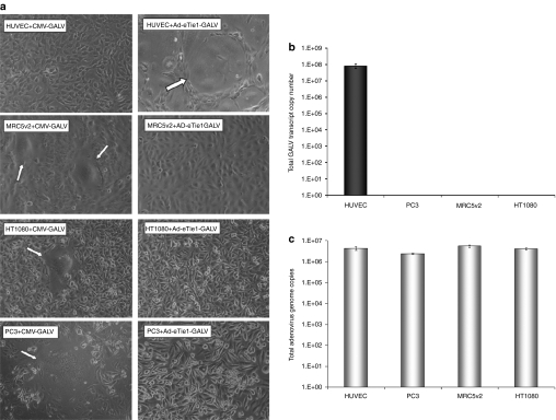
Ad-eTie1-GALV mediates extensive syncytium formation in endothelial cells but not in cells of epithelial or mesenchymal origin. (a) HUVEC, MRC5v2, HT1080, and PC-3 cells at 70% confluency were transfected with a CMV-driven GALV-expressing plasmid (left panel images) or infected with Ad-eTie1-GALV at 5,000 vp/cell (right panel images). Phase-contrast images were taken at 24 hours postinfection. Typical multinucleated syncytial structures are indicated by arrows. (b) GALV mRNA production was restricted to endothelial cells infected with Ad-eTie1-GALV. A range of cell lines including those of endothelial, epithelial, and mesenchymal origin were infected, total cellular RNA from each sample was recovered and the levels of GALV transcript were quantified by RT-QPCR. (c) Ad infectivity in each cell line was measured by harvesting cells at 24 hours postinfection with Ad-eTie1-GALV at 5,000 vp/cell, followed by DNA extraction and QPCR to quantify adenoviral genome copy numbers. Results are presented as an average of triplicates ± SD. Statistical analysis was performed using one-way ANOVA Tukey's multiple comparison test for all experimental groups. Ad, adenovirus; ANOVA, analysis of variance; CMV, cytomegalovirus; GALV, gibbon ape leukemia virus; HUVEC, human umbilical vein endothelial cell; KDR, kinase insert domain receptor; RT-QPCR, reverse-transcription real-time quantitative PCR.

