Figure 3.
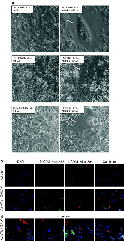
Ad-eTie1-GALV induces heterocellular endothelial–epithelial syncytium formation in vitro. Subconfluent HUVECs were infected at 5,000 vp/cell with Ad-Luc or Ad-eTie1-GALV and subsequently cocultured with uninfected PC-3, HCT116, or HEK 293 cells. (a) Phase-contrast images of syncytia (indicated by arrows) formed between HUVECs and each epithelial cell line in the presence of endothelial-specific GALV expression. (b) Fluorescence microscopy of PC-3 cells cocultured with HUVECs preinfected with Ad-Luc. (c,d) Fluorescently labeled heterocellular syncytia obtained from cocultures of PC-3 cells with HUVECs preinfected with Ad-eTie1-GALV. An anti-CD31 primary antibody was used as an endothelial-specific marker, in combination with an Alexa Fluor 594-conjugated secondary antibody. An anti-EpCAM primary antibody was used as an epithelial-specific marker, in combination with an Alexa Fluor 488-conjugated secondary antibody (see Materials and Methods section). Ad, adenovirus; CMV, cytomegalovirus; GALV, gibbon ape leukemia virus; HEK 293, human embryonic kidney 293; HUVEC, human umbilical vein endothelial cell; KDR, kinase insert domain receptor; RT-QPCR, reverse-transcription real-time quantitative PCR.

