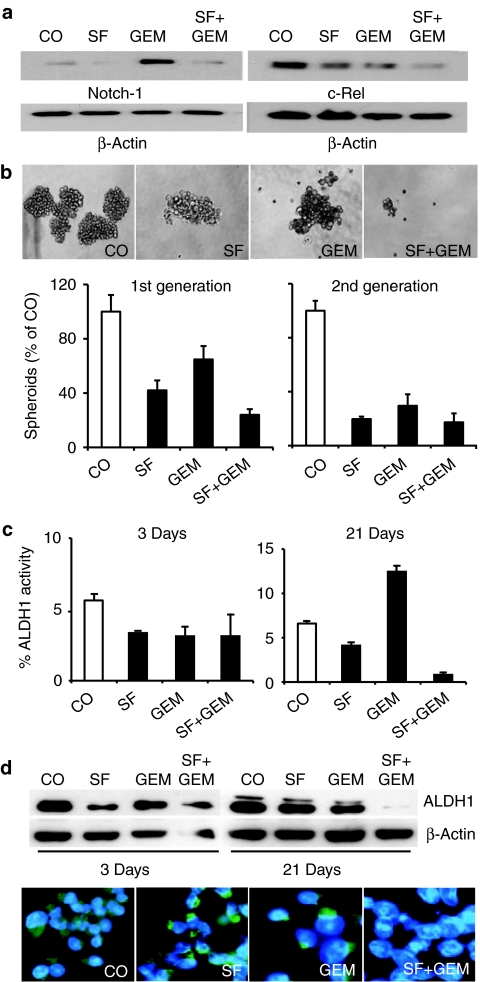
Figure 4.
Sulforaphane (SF) enhances cytotoxic drug effects to pancreatic cancer stem cell (CSC) properties. (a) Cells were treated for 48 hours with SF (5 µmol/l) and gemcitabine (GEM) (25 nmol/l) or both agents together (SF+GEM). Expression of Notch-1 and c-Rel was evaluated by western blot analysis. (b) Pancreatic CSChigh cells were seeded at clonal density in low adhesion plates for spheroid formation. Twenty-four hours later cells were treated with SF (5 µmol/l), GEM (25 nmol/l), or both agents together (SF+GEM). Spheroids were photographed at day 7 under ×100 magnification or quantified (1st generation). Thereafter, 1st generation spheroids were dissociated to single cells and equal numbers of live cells pretreatment group were re-plated. Upon spheroid formation cells were treated as described above and 3 days later spheroid formation was quantified (2nd generation). (c) Pancreatic CSChigh were treated as described above. Three or 21 days later aldehyde dehydrogenase 1 (ALDH1) activity was analyzed by flow cytometry and the percentage of ALDH1-positive cells is presented. Data are presented as mean ± SD. (d) Likewise, proteins were harvested and expression of ALDH1 protein was analyzed by western blot. Expression of β-actin served as loading control. Lower panel: Twenty-one days after treatment cells were subjected to immunofluorescence analysis for ALDH1. Randomly chosen fields were examined under ×400 magnification using a Nikon Eclipse TS100 microscope and photographs were taken.

