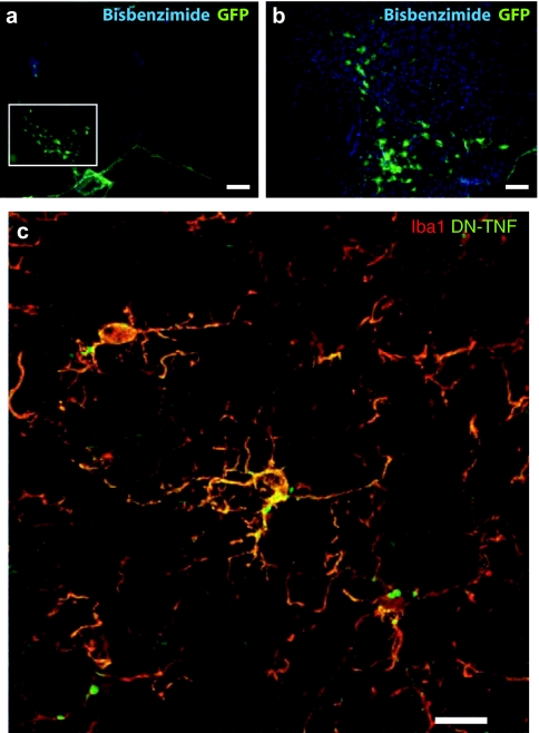
Figure 4.
Immunofluorescent localization of GFP+ cells, Iba1+ microglia, and DN-TNF protein in rats receiving DN-TNF gene transfer. (a,b) Anti-GFP immunofluorescence staining (green) in cells of glial morphology transduced in substantia nigra pars compacta (SNpc; white box). (c) Confocal microscope Z-stack of cells immunopositive for the microglial marker Iba1 (Alexa-546, red) and for hDN-TNF (Alexa-488, green) in SNpc. a, bar = 400 µm; b, bar = 200 µm; c, bar = 20 µm. DN-TNF, dominant-negative tumor necrosis factor; GFP, green fluorescent protein.

