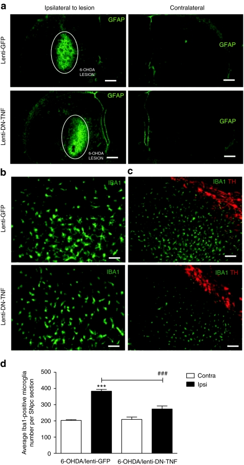
Figure 5.
Effects of nigral DN-TNF gene transfer on microgliosis and astrogliosis. (a) Activated astrocytes were identified by GFAP immunostaining (green) in the striatum. The area lesioned by the 6-OHDA neurotoxic is highlighted with a white circle. (b,c) Microglia were identified by Iba1 immunostaining (green) in the substantia nigra pars compacta (SNpc) area identified by tyrosine hydroxylase immunostaining (red). a, bar = 400 µm; b, bar = 100 µm; in c, bar = 200 µm. (d) Quantification of microglia in SNpc of 6-OHDA/lenti-GFP and 6-OHDA/lenti-DN-TNF-injected rats. Values represent the mean ± SEM of Iba1+ microglia within SNpc calculated from three separate brain sections (12 random fields/section) using threshold analysis in three animals per treatment group. Two-way analysis of variance for comparison of 6-OHDA/lenti-GFP and 6-OHDA/lenti-DN-TNF with Fisher's protected least significant difference post hoc test (###P < 0.001). DN-TNF, dominant-negative tumor necrosis factor; GFP, green fluorescent protein; 6-OHDA, 6-hydroxydopamine.

