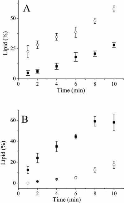
Figure 2.
Incubation of the fluorescent lipids with a cell lysate. (A) Time course of cell lysate incubated with Bodipy Fl PIP2. The solid squares and open circles are the percentage of Bodipy Fl PIP3 and peak 3 (identified in Fig 1A), respectively. (B) Time course of cell lysate incubated with Bodipy Fl PIP3. The solid squares and open circles are the percentage of Bodipy Fl PIP2 and peak 3, respectively. The data represent the average of three experiments and the error bars depict the standard deviation of the data points.

