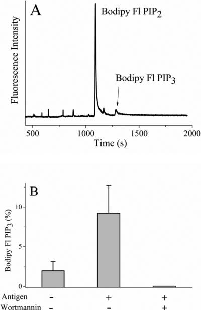
Figure 5.
Physiologic activation of RBL cells loaded with Bodipy Fl PIP2. (A) Electropherogram obtained from an RBL cell loaded with 0.5 μM Bodipy Fl PIP2:histone and then stimulated with antigen. (B) The percentage of fluorescent lipid present as Bodipy Fl PIP3 in serum starved cells (control), antigen-activated cells, and antigen-activated cells in the presence of wortmannin. The data represent the average of five experiments and the error bars depict the standard deviation of the data points.

