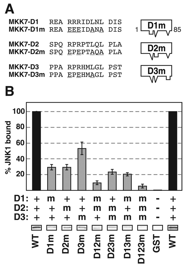
FIGURE 4. Mutation of D-sites of MKK7.
A, D1, D2, and D3 were mutated as shown, either alone or in combination, in the context of GST-MKK7-(1–85). The mutations are underlined. B, quantification of binding of MKK7 D-site mutants (40 µg) to JNK1, normalized to the binding of wild-type GST-MKK7-(1–85). Standard error bars are shown (n = 5). The key shows whether the indicated D-sites were wild-type (WT) (+), mutated (m), or absent (−). Shown below the graph are representative data bands showing the amount of 35S-JNK1 co-sedimented with the indicated GST-MKK7 mutant in a typical experiment.

