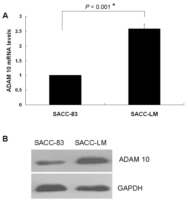
Figure 3.

ADAM 10 expression levels in SACC-83 and SACC-LM cell lines. (a) Quantitative RT-PCR showing relative ADAM 10 mRNA levels (mean ± SD) in SACC-83 cells (low metastatic potential) compared with SACC-LM cells (high metastatic potential) (*p < 0.001). (b) Western blot analysis showing ADAM 10 protein expression in SACC-83 and SACC-LM cell lines. GAPDH served as a loading control.
