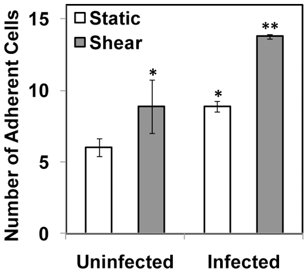
Figure 6. Monocyte adhesion to endothelial cells activated with supernatant from infected and sheared monocytes.

THP-1 monocytes were infected with mock PBS or chlamydial EB (MOI 2) for 16 h. Uninfected and infected cells were sheared for 20 min at 0 (static) or 10 dyn/cm2 (shear) using a cone-and-plate viscometer, and the supernatant was used to activate HAEC. Uninfected monocytes were perfused on top of HAEC, and the number of adherent cells was calculated in three different fields of view. The results are mean ± SD of one representative experiment performed in triplicate, and the experiments were repeated three times. The * denote statistically significant increase in the number of adherent monocytes due to shear stress or infection compared to uninfected, static control; and ** denotes statistically significant increase due to both shear stress and infection compared to all other groups (P<0.05, ANOVA).
