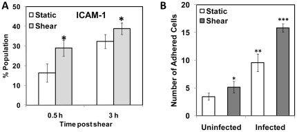
Figure 7. Effect of shear stress on ICAM-1 expression and endothelial adhesion of infected monocytes.
(A) THP-1 monocytes were infected with mock PBS or chlamydial EB (MOI 2) for 16 h. Uninfected and infected cells were sheared for 20 min at 0 (static) or 10 (shear) dyn/cm2 using a cone-and-plate viscometer, and were incubated for either 30 min or 3 h post shear. The cells were fixed, stained with FITC-conjugated ICAM-1 antibody and analyzed by flow cytometry. The * denote statistically significant increase (P<0.05, Student's t-test) in ICAM-1 expression due to shear; (B) HAECs were activated with 100 nM TNF-α and assembled in a parallel plate flow chamber. Infected monocytes were sheared either at 0 (static) or 10 (shear) dyn/cm2 for 20 min, incubated for 30 min, and then perfused through the flow chamber at 1 dyn/cm2. The number of adherent cells was counted from four different fields of view. The results are mean ± SD of one representative experiment performed in triplicate, and the experiments were repeated three times. The *, **, and *** denote statistically significant increase in the number of infected, monocytes adhered to HAEC between different groups (P<0.05, ANOVA).

