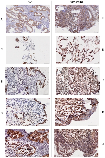
Figure 1. Endometrium cells morphology providing from luteal phase.
Revealed by immunohistochemistry with cytokeratin (KL1) and vimentin antibodies (x 6,6). A–B: before culture, C–D: after 2 days of culture without hormones and without sponge, E–F: after 2 days of culture without hormones and with sponge, G–H: after 2 days of culture with hormones (50 nmol/l estradiol +50 nmol/l progesterone) and without sponge, I–J: after 2 days of culture with hormones (50 nmol estradiol +50 nmol progesterone) and with sponge.

