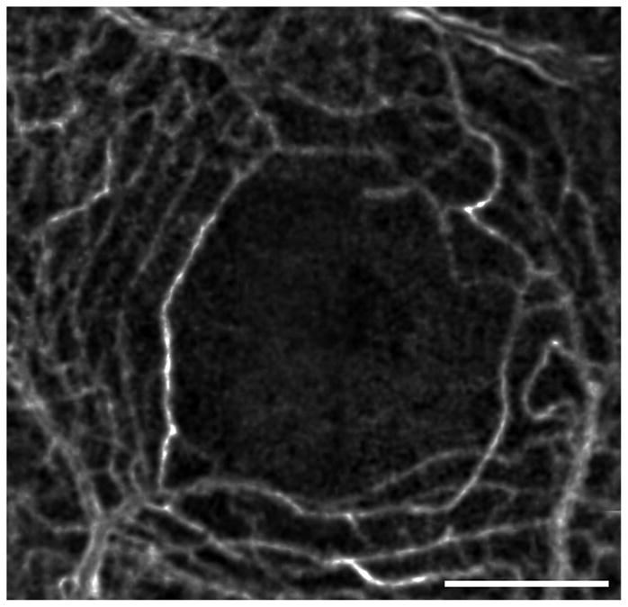
Figure 2.

Capillaries forming the edge of the FAZ in a normal eye. This image is generated by computing the motion contrast of a stabilized AOSLO video. Motion contrast images from several videos were stitched together to form this montage, showing the continuous rim of the FAZ as well as the surrounding capillary network. Scale bar is 1 degree.
