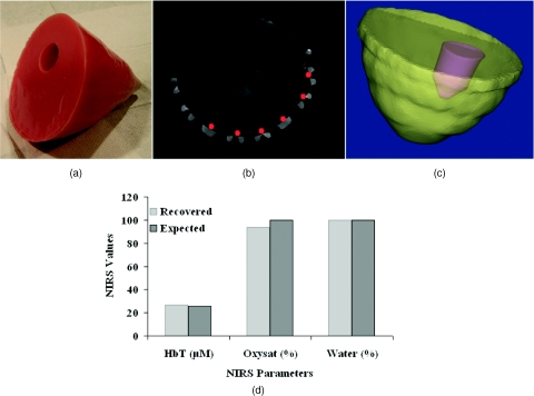
Figure 3.
(a) Breast phantom imaged in the MRI-NIR system is shown with a representative MR slice in (b) containing fiducial markers indicating the positioning of the fibers (in red) and (c) segmented background and inclusion surfaces. These surfaces were used to reconstruct the NIRS parameters of the phantom shown in (d) with comparison to expected values. Water was reconstructed with 100% accuracy in this case, because it was the maximum allowable by the reconstruction.

