Figure 1.
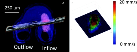
Calculating shear stress. (a) The segmented heart is indicated in blue and the blood flow in the inflow in pink. An oblique slice is positioned in an orthogonal orientation to the blood flow to calculate shear stress. (b) Velocity profile was obtained from the Doppler OCT data at the oblique cross section selected in (a) and is displayed as a surface plot.

