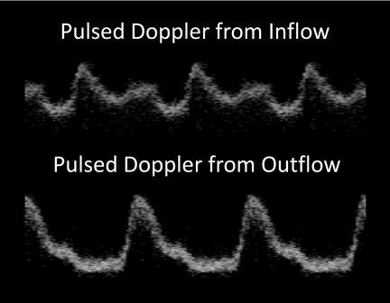
Figure 2.
Pulsed Doppler. The pulsed Doppler waveform at the inflow of the heart tube is distinctly different from the outflow waveform. The 4-D Doppler dataset enabled us to obtain pulsed Doppler traces at any location along the heart tube, which allowed easy access to multiple areas of interest. 4-D Doppler also overcomes the difficulty in selecting appropriate regions to perform M-scans in the beating heart. The top panel represents a waveform taken from the inflow of the heart tube, while the bottom panel is from the outflow of the heart tube. The inflow pulse has the two peaks characteristic of venous flow, while the outflow has a single peak characteristic of arterial flow.

