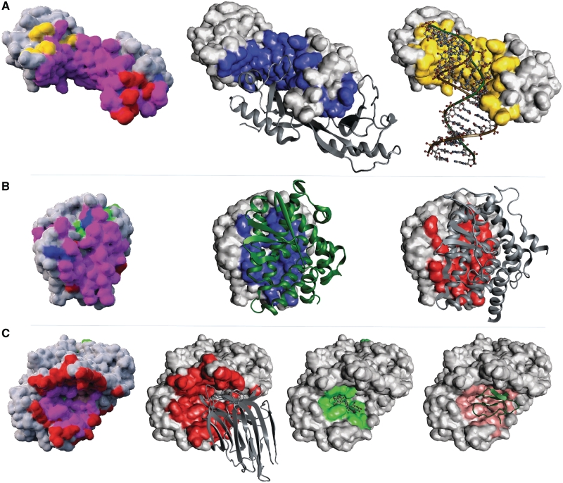
Figure 3.
Examples of proteins exhibiting polyvalent binding sites. Polyvalent binding sites are a general phenomena in molecular structures. (A) The TATA-binding protein (TBP) processed by the M-ORBIS Molecular Cartography approach starting from structure 1ais:A. (B) The Retinoid X Receptor-Alpha (RXR) cartography from the structure 1dkf:A. (C) The Pancreatic Alpha-Amylase (PAA) cartography from the structure 1dhk:A. Binding site types are represented in different colors: blue for homodimer, red for heterodimer, yellow for DNA, green for ligand and salmon for peptide. For TBP and RXR, the homodimer partner shown is extracted from structures 1d3u and 1dkf, respectively. For PAA, the heterodimeric and peptide partners are extracted from structures 1dhk and 1clv, respectively, whereas the ligand partner was extracted from 1g9h.

