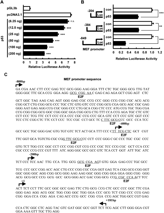
Figure 3.
p53 suppresses MEF promoter activity. (A) HCT116 p53−/− cells were transiently transfected with MEF (–849/+181 bp) promoter construct or pGL3b vector (0.2 µg) and the indicated amount of p53 plasmid or pcDNA3.1 empty vector. Luciferase activity was determined 48 h after transfection of plasmids and is expressed as fold activation over the pGL3b vector. Values are the mean ± SE of triplicate platings. ***P < 0.001 versus pcDNA3.1, determined by ANOVA with Tukey–Kramer test. n.s, not significant. (B) HCT116 p53−/− cells were transiently transfected with the indicated MEF promoter constructs (0.2 µg) and p53 plasmid (0.1 µg) or pcDNA3.1 empty vector (as control). Luciferase activity was determined 48 h after transfection of plasmids and is expressed as fold activation over the pcDNA3.1 vector (con) in each promoter construct. Values are mean ± SE of triplicate platings. **P < 0.01; ***P < 0.001 versus control, assessed by Student’s t-test. (C) The –849 bp 5′-flanking region of MEF. Nucleotide sequence of 5′-flanking region of human MEF gene is shown. The site indicated (+1) denotes the start site of the first exon. The predicted binding sites for E2F1 are marked on the sequence.

