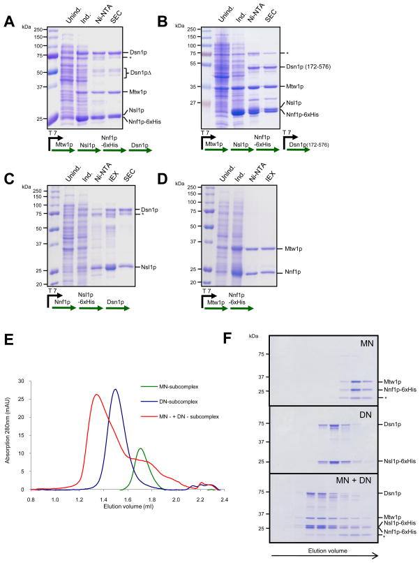
Figure 1. Reconstitution of the Mtw1 complex.
A. Expression and purification of the Mtw1 complex. The coomassie-stained gel shows different steps of the purification scheme: control extract and extract after induction with IPTG, eluate from Ni-NTA beads and purified fraction after SEC. Note the presence of degradation products of the Dsn1p subunit. B. Expression and purification of a complex with an N-terminally truncated Dsn1p subunit. C. Expression and purification of a stable Dsn1-Nsl1 heterodimer. IEX denotes anion exchange chromatorgraphy. Asterisks in A, B and C indicate a contamination with the E. coli Hsp70 chaperone D. Expression and purification of Mtw1-Nnf1 heterodimer. E. SEC runs of Dsn1p-Nsl1p and Mtw1p-Nnf1p subcomplexes (8 μM each) and of the full complex after reconstitution. F. Coomassie-stained SDS-PAGE of fractions from E. Asterisks indicate an Nnf1 truncation product.

