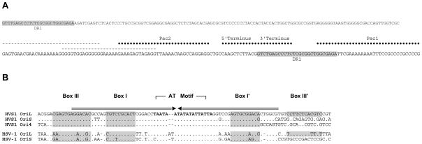
Figure 3.
Comparison of Pac1, Pac2, and replication origins of HVS1 and HSV-1. A) HVS1 Pac1 and Pac2 sites are juxtaposed by the joining of the 5′ (left; UL end) and 3′ (right; US end) termini of the genome. The dashed lines indicate possible alternate Pac2 sites. The shaded sequence identifies a repeat sequence (DR1) also found near the ends of the short repeat (RS) sequences. B) Sequences of the replication origins of HVS1 compared to the sequences of OriL and OriS sequences of HSV-1. The arrows above the alignment indicate the inverted repeat and shaded sequences identify the putative binding sites of the origin binding protein as previously defined (Hazuda et al., 1991).

