
Fig. 3.
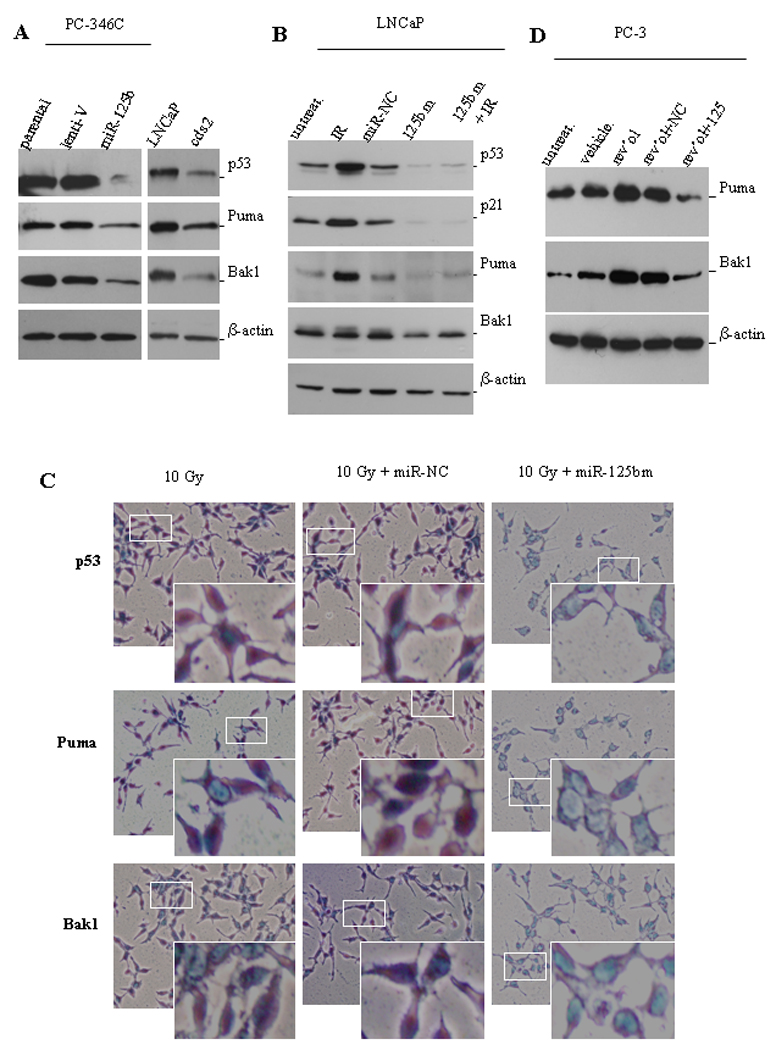
miR-125b targets pro-apoptotic genes. A) Western blot analyses of the expression levels of p53, Puma and Bak1 in lenti-miR-125b infected PC-346C cells (left panel), and in LNCaP cells grown in 10% FBS medium and in cds2 cells grown in androgen-deprived medium (right panel). Lent-V, lentiviral vector. B) Western blot analysis of the expression of p53, p21, Puma and Bak1 in miR-125bm-treated LNCaP cells. Cells were first transfected with 50 nM of chemically-modified miR-125b mimic (125bm) and 24 hours later irradiated with 10 Gys (IR). Eight hours after irradiation, cells were lysed and protein was extracted for Western blot analysis of p53, p21, Puma and Bak1. The controls include untreated cells (untreat.) and miRNA negative control (miR-NC)-treated cells. C) Detection of the expression levels of p53, Puma and Bak1 in LNCaP cells by immunostaining. LNCaP cells were grown for 24 hours on sterile slides in 100-mm Petri dishes in 10% FBS medium. Cells were first transfected with 50 nM of chemically-modified miR-125bm and 24 hours later irradiated with 10 Gys. Eight hours later, cells were fixed. p53, Puma or Bak1 were stained using specific 1st antibodies followed by a HRP-labeled 2nd antibody. Then, addition of the substrate (DAB) led to generate the brown color. The enzymatic approach shows that p53 locates mainly in nuclei, and Puma and Bak1 in cytoplasm. The white squares indicate the areas of magnified images. D) PC3 cells were transfected with 50 nM of miR-125bm (125bm) and 24 hours later treated with 100 µM resveratrol. Cells were lysed next day and protein was isolated for Western blot analysis of Puma and Bak1. Untreated cells (untreat.) and vehicle-treated cells are used as controls. β-actin is a loading control.