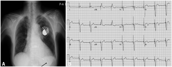
Fig. 3.
(A) Chest X-ray after pacemaker insertion. The pacemaker lead is shown at the apex of right ventricle in the X-ray (arrow), which indicates successful pacemaker insertion. (B) A 12-lead electrocardiogram after insertion of pacemaker. The electrocardiogram shows successful pacing of the pacemaker.

