Figure 2.
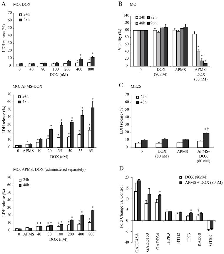
APMS-DOX delivery increases cytotoxicity in both sarcomatoid (MO) and epithelioid (ME26) MM cells in vitro. Cell lysis and viability were determined using the LDH and MTS assays, respectively. (A) LDH release by MO cells was measured under several treatment conditions including exposure to DOX alone (40–800 nM; top panel), exposure to APMS preloaded with varying concentrations of DOX (7.5×10⁶ APMS/cm2, 10–65 nM DOX equivalent; middle panel), and exposure to individual preparations of unloaded APMS (7.5×10⁶ APMS/cm2) and DOX (40–800 nM) simultaneously (bottom panel). (B) MTS viability assay confirming increased cytotoxicity of APMS-DOX (7.5×10⁶ APMS/cm2; 80 nM DOX equivalent) in MO cells. (C) LDH release by ME26 cells. For LDH, percent release was calculated based on a complete lysis induced by a positive control lysis buffer (0.09% Triton-X; data not shown). For (A, B, C), 0 = medium only control; bars represent the mean ± SEM of n = 2–4 treatment groups in duplicate or more experiments; * = p<0.05 in comparison to medium alone at same time point; † = p<0.05 in comparison to DOX alone group. (D) PCR array experiments reveal alteration in several genes related to DNA damage and repair in sarcomatoid (MO) MM cells treated with DOX (80 nM) or APMS-DOX (7.5×10⁶ APMS/cm2; 80 nM DOX equivalent) for 24 h. Upregulation of 6 genes and downregulation of 1 gene was observed following treatment with DOX or APMS-DOX compared to cells treated with medium alone. No significant gene expression alterations were observed with APMS alone. Only gene changes ≥3-fold are provided. * = p<0.05 in comparison to DOX alone group. † = not significant compared to medium control group. Bars represent the mean +/− SEM of n = 3 plates/group.

