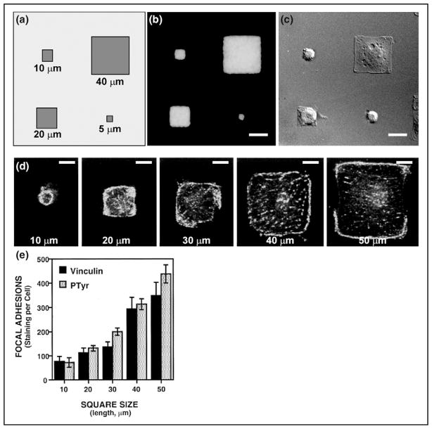
Figure 4.
Cell shape-dependent control of focal adhesion formation on micropatterned adhesive islands of different size coated with fibronectin. (A) Diagram of an assortment of square adhesive islands with sides ranging from 10 to 50 μm in length that were micropatterned onto substrates using microcontact printing. (B) An immunofluorescence micrograph showing staining for adsorbed fibronectin selectively limited to the square islands. (C) A differential interference contrast micrograph of bovine capillary endothelial cells cultured on different sized, square fibronectin islands. (D) Fluorescent confocal micrographs of individual, vinculin-labeled cells cultured on square islands of different sizes (lengths of sides are indicated). (E) Quantitation of total vinculin and total phosphotyrosine labeling per cell, for cells cultured on different sized squares. Over 30 cells per condition were averaged; error bars indicate standard error of the mean (The image is kindly provided by Dr. Ingber, and reproduced with permission from [43]).

