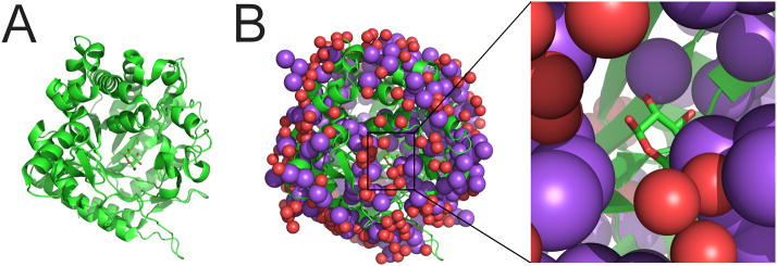
Figure 3.
Crystal structure of β-glucosidase (PDB code 3FJ0) [76]. (A) Overall structure shown in ribbon representation, with a reaction intermediate shown in stick representation. (B) The structure has an unusually large number of Na+ ions (purple spheres). Water molecules are marked as red spheres. The inset shows the binding site of the reaction intermediate in greater detail. The automatic procedures described in [37,38] do improve the R factors, but do not correct the misidentification of waters as sodium ions: after automatic rerefinement, the resulting structure contains the same erroneous number of Na+ atoms (252).

