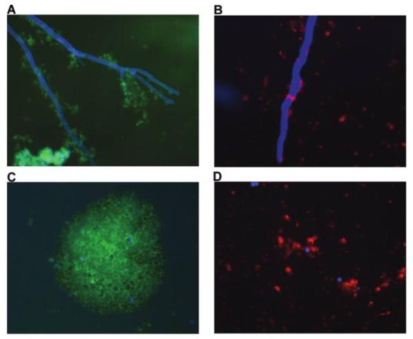
Figure 1.

Representative fluorescence photomicrographs of platelet adherence (A, C) and activation (B, D) after contact with either hyphae or conidia of Zygomycetes, which were stained with calcofluor white (blue fluorescence). Platelets adhered to hyphae (A) and conidia (C) of zygomycetes and were visualized by monoclonal anti CD-42b–fluorescein isothicyanate conjugated antibody (green fluorescence). Platelets became activated after contact with either hyphae (B) or conidia (D) of zygomycetes as shown by monoclonal anti-CD62P-PE antibody (red fluorescence), which is presented exclusively on activated platelets.
