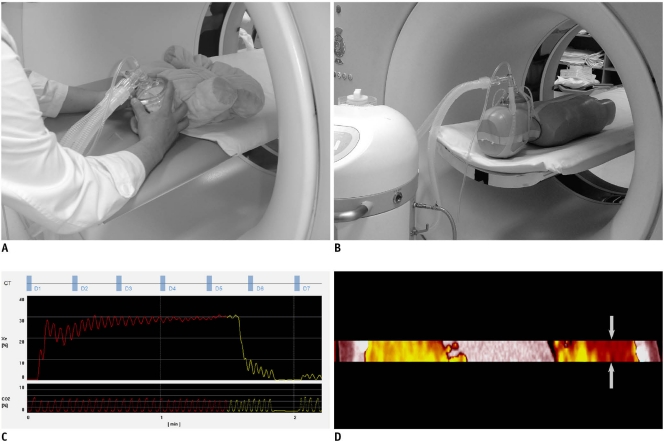
Fig. 1.
Imaging techniques of xenon-enhanced dynamic dual-energy CT.
A. For young, sedated child (doll for demonstration), face mask is fitted with hands during CT examination. B. In older, cooperative child (5-year-old anthropomorphic phantom for demonstration), face mask with elastic straps is used. C. Diagram demonstrates dynamic dual-energy spiral CT scan protocol (D1-D7) during xenon wash-in (red) and wash-out (yellow) periods. Baseline scan (D1) is followed by wash-in (D2-D5) and wash-out scans (D6 and D7). Interscan delay of dynamic scans is set to 10-20 sec. Exhaled xenon concentration reaches 30% at end of wash-in period. Real-time monitoring of exhaled xenon and carbon dioxide concentrations (%) during CT examination is also shown. D. Dual-energy spiral scan is acquired in middle of lung lesion with shortest longitudinal coverage, approximately 2.6-3.0 cm, during free-breathing. Ventilation defect (arrows) is noted in left lung when right lung shows maximal xenon enhancement.

