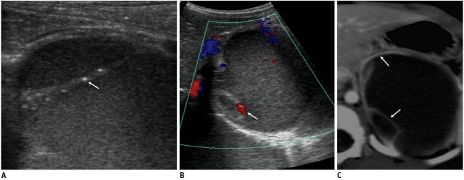
Fig. 1.
Congenital cystic neuroblastoma in 61-day-old girl.
A. US shows internal turbidity and tiny calcifications (arrow) in internal septum. B. Color Doppler US demonstrates septal vascularity (arrow). C. Contrast-enhanced CT scan shows enhancement of internal septum and outer wall of cyst (arrows).

