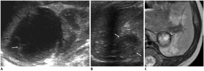
Fig. 5.
Evolution of cystic neuroblastoma in 63-day-old boy.
A. Initial US shows left adrenal cystic mass (arrows) with internal septation (white arrow). B, C. Cyst (white arrows) became smaller and more complex solid cystic masses on US (B) and T2-weighted MR (C) images obtained after one and two months, respectively. Dark signal intensity rim may suggest previous hemorrhage (arrows).

