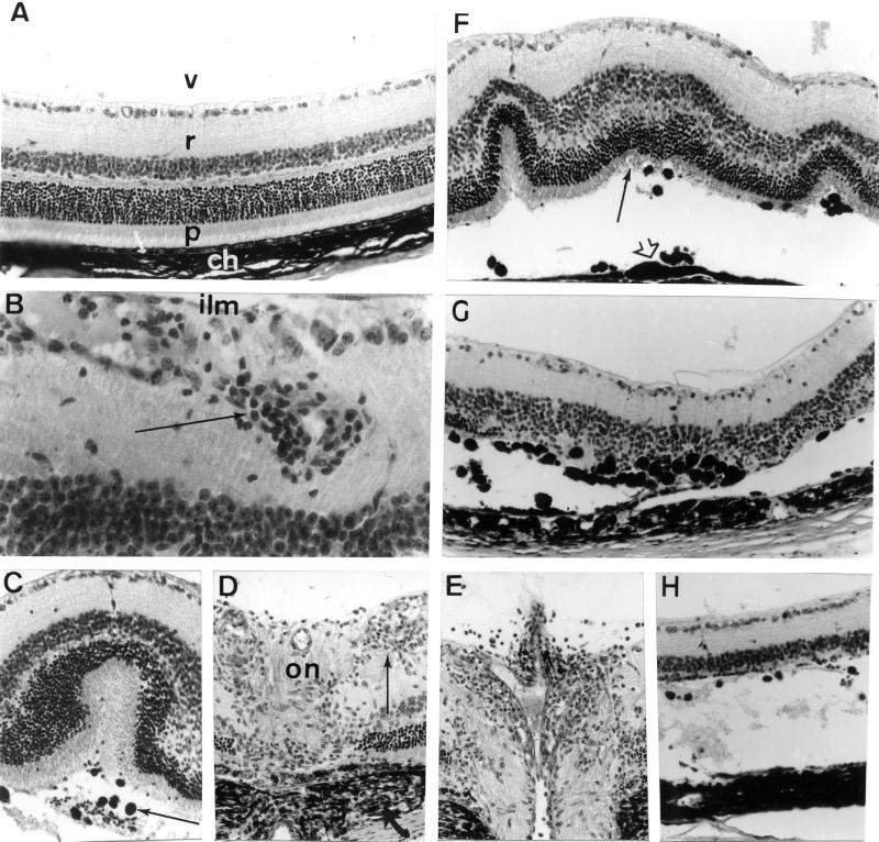
Figure 2.
Ocular histopathology of the posterior segment of the eye of aging (12 months) A29-negative and A29-positive mice. (A) Posterior segment of an eye from an A29-negative mouse: v, vitreous; r, retina; p, photoreceptor outer segments of visual cells; arrow, retinal pigmented epithelium; ch, choroid. (B–H) Posterior segments of eyes from A29-positive mice. (B) Retinal vasculitis and perivasculitis with infiltrating inflammatory cells in the vicinity of retinal vessels located near the internal limiting membrane (ilm) of the retina (arrow). (C) Retinal fold with serous exudate and retinal pigmented epithelium migration (arrow) observed in the subretinal space of the retina. (D and E) Inflammatory cell infiltration (thin arrow) at the optic nerve head (on) with choroidal inflammation adjacent to the optic disk (arrow, D) and papillitis (E). (F) Several retinal folds with slightly disorganized photoreceptor cell layer (arrow), retinal pigmented epithelium showing aspects of in situ proliferation (arrowhead), and mobilization in the subretinal space. (G and H) Two aspects of retinal and retinal pigmented epithelium pathology with partial destruction of the photoreceptor cell layer and intense retinal pigmented epithelium migration in the retina (G) to almost complete destruction of the photoreceptor cell layer and subretinal serous detachment (H). Paraffin-embedded were sections stained with hematoxylin, eosin, and safran solutions. (Magnification: A, ×800; B, ×1350; C, ×600; D, ×700; E, ×650; F, ×600; G, ×650; H, ×650.)

