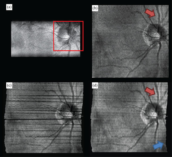
Fig. 6.

Motion compensated interlaced SECSLO-SDOCT of in vivo human retina. (a) A region-of-interest (red box) in a series of 100 5 x 3 mm2 (lateral x spectral) raw SECSLO fundus images were co-registered using an affine transformation. The resulting transformation matrices were used to compensate for bulk motion in an SDOCT volume acquired interlaced with the SECSLO fundus images. (b) 5 x 5 mm2 SDOCT SVP without motion compensation. (c) Maximum intensity projection of SDOCT SVP data following correction for bulk motion (black areas represent regions of data never acquired due to patient motion), and (d) interpolated motion compensated SVP demonstrating the utility of real-time SECSLO fundus imaging as a method for motion tracking and image registration of SDOCT B-scans. In these data sets, discrete lateral discontinuities, as a result of saccades, were compensated using motion tracking (red arrow). Furthermore, accumulated lateral drifts during imaging, with total displacements of several hundred microns (Fig. 5), resulted in misrepresentations of the structural morphology in the raw SDOCT volume as evidenced by the distorted borders of the SVP (blue arrow), which were also compensated using the co-registered SECSLO fundus images.
