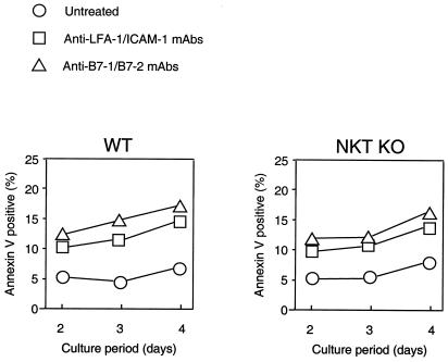
Figure 3.
Quantitative analysis of apoptosis of alloreactive responder cells in vitro. Splenocytes (2 × 10⁶ cells) from WT (Left) or NKT KO (Right) B6 mice were cocultured with mitomycin C-treated BALB/c splenocytes (2 × 10⁶ cells) in 6-well flat-bottomed plates in the presence or absence of 1 μg/ml each of anti-LFA-1/ICAM-1 or anti-B7–1/B7–2 mAbs. After 2–4 days, the cells were collected and stained with FITC-labeled anti-H-2Kb mAb and phycoerythrin-labeled annexin V. The proportion of annexin V-positive apoptotic cells in the H-2Kb-positive responder cells was determined by fluorescence-activated cell sorter (FACS) Calibur. Data represent mean of four wells. Similar results were obtained in three independent experiments.

