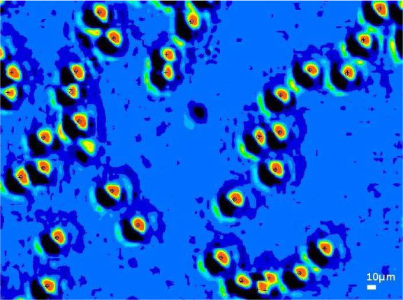
Fig. 10.

Lensless imaging of 1µm beads in a thin wetting film. The image is upsampled by a factor 9. Intensities are indicated in 16 colors on a linear scale. Overplotted in black are the centroid of the fluorescence detection (cross) and lensless imaging position (circle).
