Fig. 3.

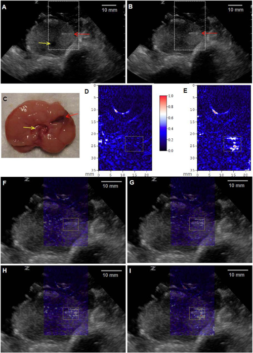
In vivo PAT and US dual-modality imaging of a canine prostate with a subsurface lesion induced. Gray scale US images of the prostate acquired (A) before and (B) after the generation of the lesion (0.1 mL of injected blood). The red arrow indicates the plastic canula inserted into the prostate for the injection of blood. The yellow indicates the urethra. (C) Anatomical photograph of the imaged cross-section in the prostate with the lesion marked. PAT images of the prostate right lobe acquired (D) before and (E) after the generation of the lesion (0.1 mL of injected blood), where the image intensities are demonstrated with an image color bar. The PAT imaging plane is the same as that in US images, where the reconstructed area is indicated with the dashed rectangles in the US images in (A) and (B). In (F)-(I), PAT images are superimposed on the US image in (B). (F) was acquired before the generation of the lesion; (G), (H) and (I) were acquired after three injections of blood, where the total blood volumes in the lesion were 0.025 mL, 0.05 mL and 0.1 mL, respectively. The area of the lesion is marked with a dotted rectangle in each image.
