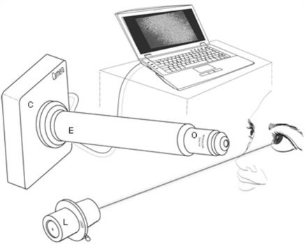
Fig. 1.

The schematic of the FASIC setup is shown here. A 635nm low power (<1mw) laser is illuminated onto the cornea in order to measure the pre-corneal tear film thickness. The reflecting and scattering light is captured by a cMOS camera that is positioned in an angle with respect to the incoming light. A stack of 256X256 pixel images are streamed to a computer through a firewire cable for further analysis.
