
Fig. 5.
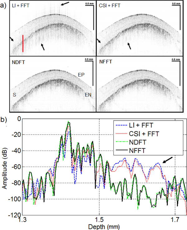
(a) Corneal images obtained from different processing techniques. The arrows indicate the location of the image artifacts. EP, epithelium; S, stroma; EN, endothelium. (b) Representative part of an A-line located at the solid line in the corneal image. NFFT produced peaks with higher intensity as a result of the improved sensitivity fall-off. LI, Linear interpolation; CSI, cubic spline interpolation; NDFT, non-unifrom discrete Fourier transform; NFFT, non-uniform fast Fourier transform.