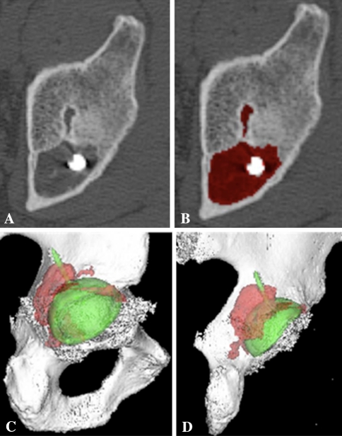
Fig. 4A–D.
(A) Preprocessing axial CT cut of a lytic lesion believed to be osteolysis because there was no preexisting cyst in this area on the immediate postoperative radiograph. (B) Mapping of the lesion on the axial view is shown. (C) The preprocessing axial view was mapped to create a three-dimensional model of the lytic lesion. (D) The axial view was mapped to create a three-dimensional model of the lytic lesion.

