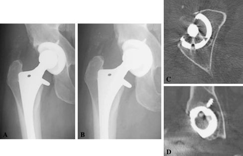
Fig. 5A–D.
Representative images are shown from the single case of osteolysis in a patient with a highly crosslinked polyethylene liner. (A) An AP radiograph from the immediate postoperative period is shown. (B) An AP radiograph from the most recent followup is shown. (C) A representative axial slice of the CT scan is shown for this patient. (D) A representative coronal slice of the CT scan for this patient is shown.

