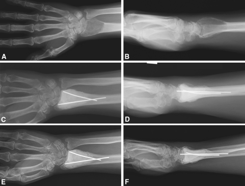
Fig. 1A–F.
(A) AP and (B) lateral views are shown of a GCT of the distal radius at diagnosis. (C) AP and (D) lateral radiographs obtained 6 months after intralesional surgery with polymethylmethacrylate filling are shown. (E) AP and (F) lateral radiographs obtained at the 3-year followup show no signs of recurrence.

