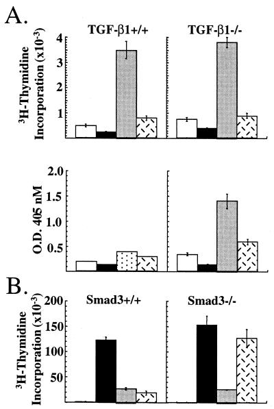
Figure 3.
Inhibition of TGF-β1−/− and smad3−/− T cells by CTLA-4. (A) Purified T cells from TGF-β1+/+ (Left) and TGF-β1−/− (Right) were cultured with latex microspheres coated with antibody to CD3 alone (open bars), anti-CD3/anti-CTLA-4 antibody (solid bars), anti-CD3/anti-CD28 antibody (shaded bars), or anti-CD3/anti-CD28/anti-CTLA-4 antibody (hatched bars). Proliferation (Upper) and IFN-γ secretion (Lower) were assessed at 72 h. (B) Purified T cells from smad3+/+ (Left) and smad3−/− (Right) mice were stimulated with soluble control Ig (open bars), anti-CD3/anti-CD28 antibody (solid bars), anti-CD3/anti-CD28/anti-CTLA-4 antibody (shaded bars), and anti-CD3/anti-CD28 antibody plus rHuTGF-β1 (2.5 ng/ml) (hatched bars). Proliferation was assessed at 72 h.

