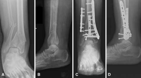
Fig. 1A–D.
(A) AP and (B) lateral radiographs show a comminuted intraarticular pilon fracture with concurrent fibular fracture and valgus malalignment. (C) AP and (D) lateral radiographs after open reduction and internal fixation with correction of limb alignment show near anatomic restoration of the articular surface.

