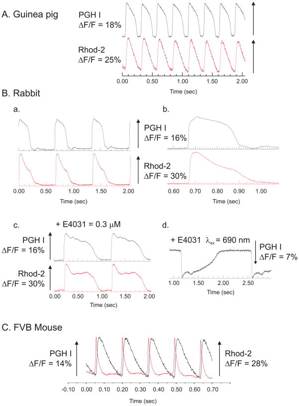
Figure 7. Simultaneous recordings of APs and intracellular Ca2+ transients.
APs and Cai transients were mapped simultaneously from Langendorff perfused hearts that were stained with both PGH I and Rhod-2 by focusing images of the heart on 2 16×16 elements photodiode arrays (Choi et al., 2002; Choi & Salama, 2000).
A: Simultaneous AP and Cai recordings from a guinea pig heart. The superposition of APs and Cai transients recorded from 2 diodes, one on the ‘voltage’ array the other diode from the ‘calcium’ array where both diodes are aligned to view the same 0.9 × 0.9 mm2 region of epicardium.
B: Simultaneous AP and Cai recordings from a rabbit heart
APs and Cai transients as in (A), except that the recordings were made from a pre-pubertal female rabbit heart at slow (a) and fast (b) sweep speed. The addition of E4031 (0.5 μM), a known blocker of IKr (the fast component of the delayed rectifying K+ current) caused a marked prolongation of the AP duration which was readily measured with PGH I (traces c). Longer excitation wavelength (690 instead of 540 nm) caused the inversion of ΔF and an inverted AP (c).
C. Simultaneous AP and Cai recordings for an FVB mouse heart
Langendorff perfused mouse hearts were stained with a PGH I stock solution (2 mM in Tyrode’s solution at pH 6.0) and Rhod-2/AM. The superposition of AP and Cai traces from 2 diodes viewing the same region of myocardium (250 × 250 μm2) show that for mouse ventricular muscle the Cai transients are considerably longer than the AP.

