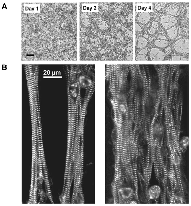
Figure 2. Fiber appearance.

(A) Fiber structure developed during the first 3–4 days after which relatively few morphological changes occurred. Bright field images of live unstained samples from the same preparation during Days 1, 2, and 4 are shown. (B) The myocytes inside the fibers assume aligned orientation, which resembles intact cardiac tissue. Images show thin and thick fibers stained for the sarcomeric protein α-actinin.
