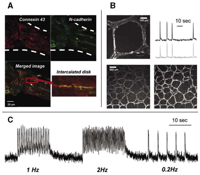
Figure 3. Accessibility of fibers to live and fixed tissue probes.

(A) Example of thick cardiac fiber stained with connexin 43 (red) and N-cadherin antibodies (green). An enlarged inset from the merged image illustrates the formation of an intercalated disk, a signature of cardiac muscle development. (B) The top left image shows highly organized fibers with myocytes aligned along each fiber segment (20x objective). The top right image shows calcium transients recorded from a net of spontaneously beating medium-sized cardiac fibers loaded with calcium-sensitive dye Fluo-4. The bottom two frames (4x objective) show the passage of the excitation wave through the fiber net (see Supplementary Movie S4). (C) Calcium transients from macrofibers paced by external electrodes with three trains of stimuli at 1,2, and 0.2Hz frequencies. Elevated levels of diastolic calcium seen in 1 and 2Hz traces are due to incomplete re-uptake of cytosolic calcium, resulting from fast pacing at room temperature.
