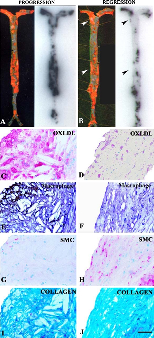
Fig. 4.
En face preparation of Sudan-stained mouse aortas (left) and corresponding autoradiographs (right) showing representative examples of intravenously injected 125I-MDA2 distribution in the progression (a) and regression (b) groups. Arrowheads depict Sudan-stained areas with diminished 125I-MDA2 uptake. Panels c–j represent examples of immunostained atherosclerotic lesions from LDLR−/− mice from the progression (c, e, g, i) and regression (d, f, h, j) groups. Immunostaining was performed for OxLDL, macrophages, smooth muscle cells (SMCs), and collagen. OxLDL and SMCs appear pink/purple, macrophages black, and collagen bright blue. Scale bar = 50 μm. (Reprinted with permission from Torzewski et al. [11••])

