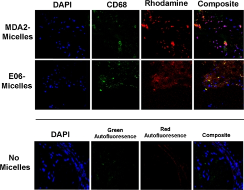
Fig. 7.
Confocal microscopy (63×) of apoE−/− mouse aortic atherosclerotic plaque following the administration of MDA2-micelles and E06-micelles demonstrating nuclei (blue), macrophages (green), and micelles (red). The lower panels represent an atherosclerotic plaque from an apoE−/− mouse not injected with micelles and stained with DAPI, showing the absence of significant autofluorescence (40×). (Reprinted with permission from Briley-Saebo et al. [22••])

