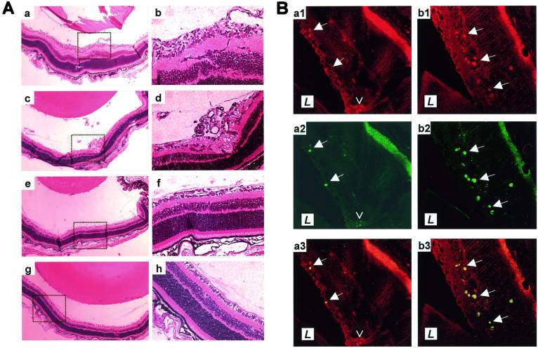
Figure 1.
In situ angiogenesis and apoptosis of endothelial cells in PEDF-treated retinas. (A) Examples shown at low (Left) and high (Right) power of retinas of mice exposed to hyperoxia and subsequently treated with vehicle (Aa–Ad), with 11.2 μg/day PEDF (Ae and Af) or with 22.4 μg/day PEDF (Ag and Ah). (B) Sections of retinas from mice treated with vehicle (Ba) or with 11.2 μg/day PEDF (Bb) were stained with PECAM-1 to detect endothelial cells (Ba1 and Bb1) and with TUNEL to detect apoptotic cells (Ba2 and Bb2), and the images merged (Ba3 and Bb3). L indicates the position of the lens. Solid arrows are reference points within each retina. The open arrowhead in panel Ba1–Ba3 points to a vascular tuft of predominantly TUNEL-negative endothelial cells. Note that the retinal pigment epithelial layer autofluoresces.

