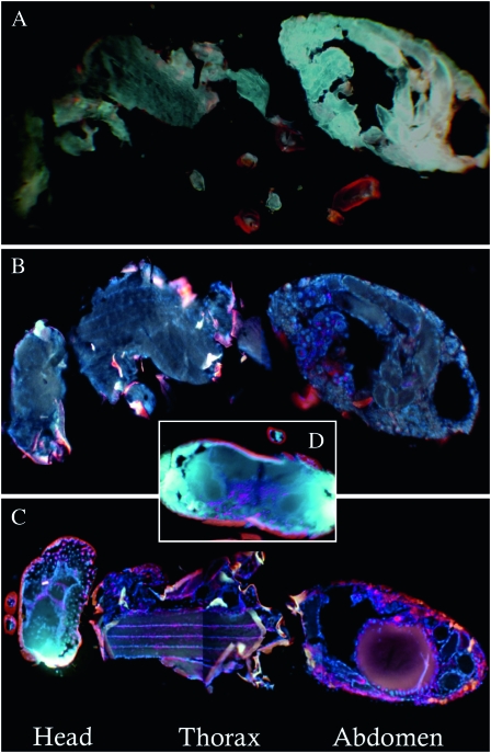
Figure 2.—
Fluorescent in situ hybridizations of W. pipientis in adults of N. vitripennis and N. giraulti. Comparisons of 30-μm cross-sections from adult females of (A) unstained N. vitripennis 12.1 (control for autofluorescence), (B) stained N. vitripennis 12.1, (C) stained N. giraulti IntG12.1, and (D) a stained N. giraulti IntG12.1 head. Strong W. pipientis signals (red) are observed in IntG12.1 in the head, the flight mussel tissues in the thorax, and the abdomen. Images B–D are counterstained with DAPI (blue) and are presented as composite images from the same individual to enhance resolution of the W. pipientis.

